Upper Gastrointestinal Ulcer Model Endoscopic Hemostasis Simulator

This product was developed through a joint research collaboration between Tohoku University and Denka Company Limited as a simulator capable of replicating bleeding to enhance hemostasis techniques. Designed for both beginners and experts, it allows training using actual endoscopes and devices. The independently developed soft material faithfully reproduces the realistic tactile sensation of the treatment area. Additionally, pulsatile bleeding (Forrest Ia) is simulated through a syringe. The Upper Gastrointestinal Tract model, into which an actual endoscope can be inserted, is used by attaching either an ulcer model for clips or an ulcer model for coagulation grasper.
Background of Development
Among gastrointestinal bleeding, particularly in cases of profuse bleeding, it is life-threatening.Therefore, there is a need to improve the skills of hemostatic techniques. However, there was no medical training model available that could replicate bleeding.
Development Concept
- Enables learning of endoscopic therapy using actual endoscopes and devices.
- Provides medical training opportunities for beginners to experts.
- Constructed with specific soft resin that can be stored for long periods.
Features
- Reproduces realistic tactile sensations of the procedural area with proprietary specific soft resin
- Reproduces pulsatile bleeding (Forrest Ia) through syringe manipulation.
- Technical levels can be adjusted by changing the attachment positions.
- Allows practice with four blood vessels per ulcer.
- Selective bleeding can be controlled by a three-way stopcock.

Ulcer model for clips

Resembling the human gastrointestinal mucosa in elasticity and color tone

Grasping the ulcer with a hemostatic clip
Ulcer model for coagulation grasper

Compatible with integrated bipolar plates for coagulation

Grasping the ulcer and coagulating with a coagulation grasper
High Reproducibility and Simple Setup
- Place the ulcer model inside the upper gastrointestinal tract part and connect the blood vessels to the three-way stopcock for a complete setup.
- The setup can be easily done, making it ideal for remote guidance.
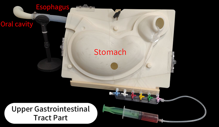
By providing lumens from the oral cavity to the treatment site, the entire operation process can be learned.The simulator can be easily disassembled for washing, and the lumen part can be repeatedly used.
Retrospective larning
The ulcer model can be peeled off from the upper gastrointestinal tract part after the practice to confirm the clip positions and application of the coagulation grasper, facilitating the understandings of aquisition levels.

Related Videos
Behind the Product Development Story
How to Assemble the Upper Gastrointestinal Ulcer Model
